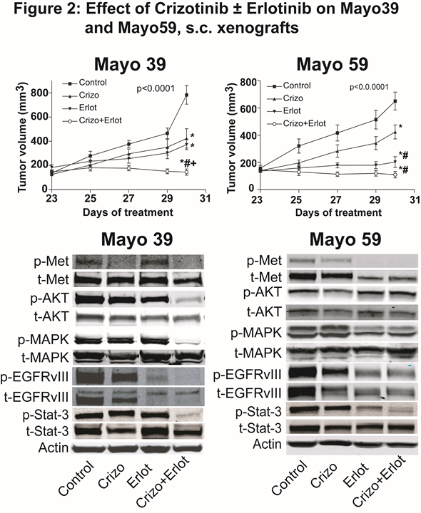
Introduction:\r\nThey are implicated in the malignant progression of glioblastoma via distinct and overlapping oncogenic downstream signaling pathways. EGFRvIII, a constitutively activated EGFR deletion mutant variant, leads to increased tumor growth and diminishes the tumor growth response to HGF:c-Met pathway inhibitor therapy. Activation of the c-Met pathway diminishes the tumor growth response to EGFR pathway inhibitors EGFRvIII and c-Met pathway inhibitors synergize to inhibit tumor growth in isogenic GBM cell lines engineered to express EGFRvIII. Despite targeting RTK signaling in glioblastoma multiforme, a subpopulation of stem-like tumor-propagating cells can persist to replenish the tumor cell population leading to tumor recurrence.\r\n\r\nMethods:\r\nCell Culture: Mayo 39 and Mayo 59 xenograft lines were purchased from the Mayo clinic, as live subcutaneous (s.c.) xenograft-bearing mice. Xenografts were maintained through serial subcutaneous re-implantation in immunodeficient mice (Athymic NCr-nu/nu, NCI). Human clinical specimens were obtained from the Johns Hopkins Neurosurgical operating suites. S.C. Xenografts: Mayo 39 and Mayo 59 tumor xenograft lines serially passaged in nude mice were used to generate s.c. xenografts for experimental studies. When tumors were approximately 150 mm3, mice were randomly divided into 4 groups: 1) control vehicle, 2) Crizotinib, 3) Erlotinib, or 4) Crizotinib + Erlotinib, and received the indicated dose of Crizotinib (40 mg/kg/d, gavage), Erlotinib (100 mg/kg/d, gavage) or the combination of both for 7 days. Control animals received corresponding amounts of DMSO. Tumor sizes were measured every alternate day and tumor volumes were estimated by measuring two dimensions [length (a) and width (b)] and calculated tumor volumes (V) using the equation: V=ab2/2. At the termination of each experiment, tumors were excised and harvested. Antibodies: Antibodies were obtained from Santa Cruz Biotechnology, Ventana Medical Systems, and Sigma. Immunohistochemistry: Paraffin sections were incubated with anti-cleaved caspase-3, antilaminin or anti-Ki-67 primary anti-body followed by appropriate biotinylated-conjugated secondary antibodies. Tumor cell proliferation, apoptotic, and blood vessel density indices were determined by computer assisted quantification using Image J Software. Immunoblot Analyses: Immunoblotting was performed according to standard procedures. For immunoblot analyses, proteins were electrophoretically transferred to a nitrocellulose membrane. Membranes were incubated with primary antibodies, washed, incubated with the secondary antibody, and washed. Proteins were detected and quantified using the Odyssey Infrared Imager (LI-COR Biosciences). Βeta-Actin was used as a loading control. Neurosphere (NS) formation: Control and treated tumors were harvested to isolate NS. NS were embedded in agarose, stained with Wright stain and counted by computer assisted image analysis. Statistical analysis: POne-way ANOVA followed by the Tukey Multiple comparison test using Prism (Graph Pad Software Inc.). p-value < 0.05 was deemed statistically significant.\r\n\r\nConclusion: \r\nMayo 39 and Mayo 59 xenografts express high levels of phospho-EGFR-VIII and phospho-c-MET, making Mayo 39 and Mayo 59 glioma cells excellent models to assess the combined effects of anti-EGFR and anti c- Met therapy. Crizotinib (c-Met pathway inhibitor) and Erlotinib (EGFR pathway inhibitor) in combination significantly inhibit tumor growth, downstream signaling molecules, and neurosphere growth in primary GBM subcutaneous xenografts The expression of stem cell markers Nestin, Musashi, Olig 2 and Sox2 were significantly down-regulated by c- Met inhibition No additive effect was seen by co-treatment with Erlotinib. Targeting these two parallel pathways provides substantial anti-tumor activity in glioblastoma models.\r\n\r\nReferences:\r\n• Engelman JA, Zejnullahu K, Mitsudomi T, Song Y, Hyland C, Park JO, et al: MET amplification leads to gefitinib resistance in lung cancer by activating ERBB3 signaling. Science 316:1039-1043, 2007\r\n• Lal B, Goodwin CR, Sang Y, Foss CA, Cornet K, Muzamil S, et al: EGFRvIII and c-Met pathway inhibitors synergize against PTEN-null/EGFRvIII+ glioblastoma xenografts. Mol Cancer Ther 8:1751-1760, 2009\r\n• Lal B, Xia S, Abounader R, Laterra J: Targeting the c-Met pathway potentiates glioblastoma responses to gamma-radiation. Clin Cancer Res 11:4479-4486, 2005\r\n• Stommel JM, Kimmelman AC, Ying H, Nabioullin R, Ponugoti AH, Wiedemeyer R, et al: Coactivation of receptor tyrosine kinases affects the response of tumor cells to targeted therapies. Science 318:287-290, 2007\r\n• Xu H, Stabile LP, Gubish CT, Gooding WE, Grandis JR, Siegfried JM: Dual blockade of EGFR and c-Met abrogates redundant signaling and proliferation in head and neck carcinoma cells. Clin Cancer Res 17:4425-4438, 2011\r\n